
ABiR

Prof. Daniela Massi (Section of Pathological Anatomy, DSS - Scientific Responsible)
Dott. Francesco De Logu (Section of Clinical Pharmacology and Oncology, DSS – Laboratory Coordinator)
The ABiR laboratory is located at the Centro Polivalente il Cubo 27a (first floor, north wing, room 001/40), Viale Pieraccini 6, Florence.The innovative equipment in the laboratory is dedicated to the multiparametric analysis of
tissues for the evaluation of the expression of biomarkers in tissues with the highest
accuracy and precision, in order to obtain qualitative and quantitative information on the
distribution of tumor cells and their interaction with cells of the immune system in space
and time. ABiR carries out scientific research activities funded by the 5x1000 program of
the Italian Association for Cancer Research (AIRC) for activities related to oncological
pathology, particularly melanoma (Prof. Daniela Massi), and the European Research
Council (ERC) under the European Union’s Horizon 2020 Programme for Research and
Innovation (Prof. Pierangelo Geppetti) for activities related to the role of Schwann cells in
the development and maintenance of chronic pain.
The equipment includes:
- Preparation station for histological samples, from tissue sampling to paraffin embedding
and cryopreserved tissue. - Leica Microtome and Cryostat for sample cutting and slide preparation.
- ROCHE Automatic Immunostainer (Ventana Discovery Ultra), for automated slide
staining, with the possibility of setting protocols for IHC Multiplex in Bright Field and
Fluorescence and RNA scope. - Three Slide Scanners (Leica AT2, Leica CS2, and Menarini D-Sight) that can perform
high-speed digital scans (up to 400 slides per day) (Whole Slide Imaging). - Zeiss Axio Imager 2 microscope equipped with Apotome2 (structured light), which allows
the acquisition of multiple planes on the Z-axis with high image quality and absence of
noise, and support for 8 slides capable of performing high magnification scans in
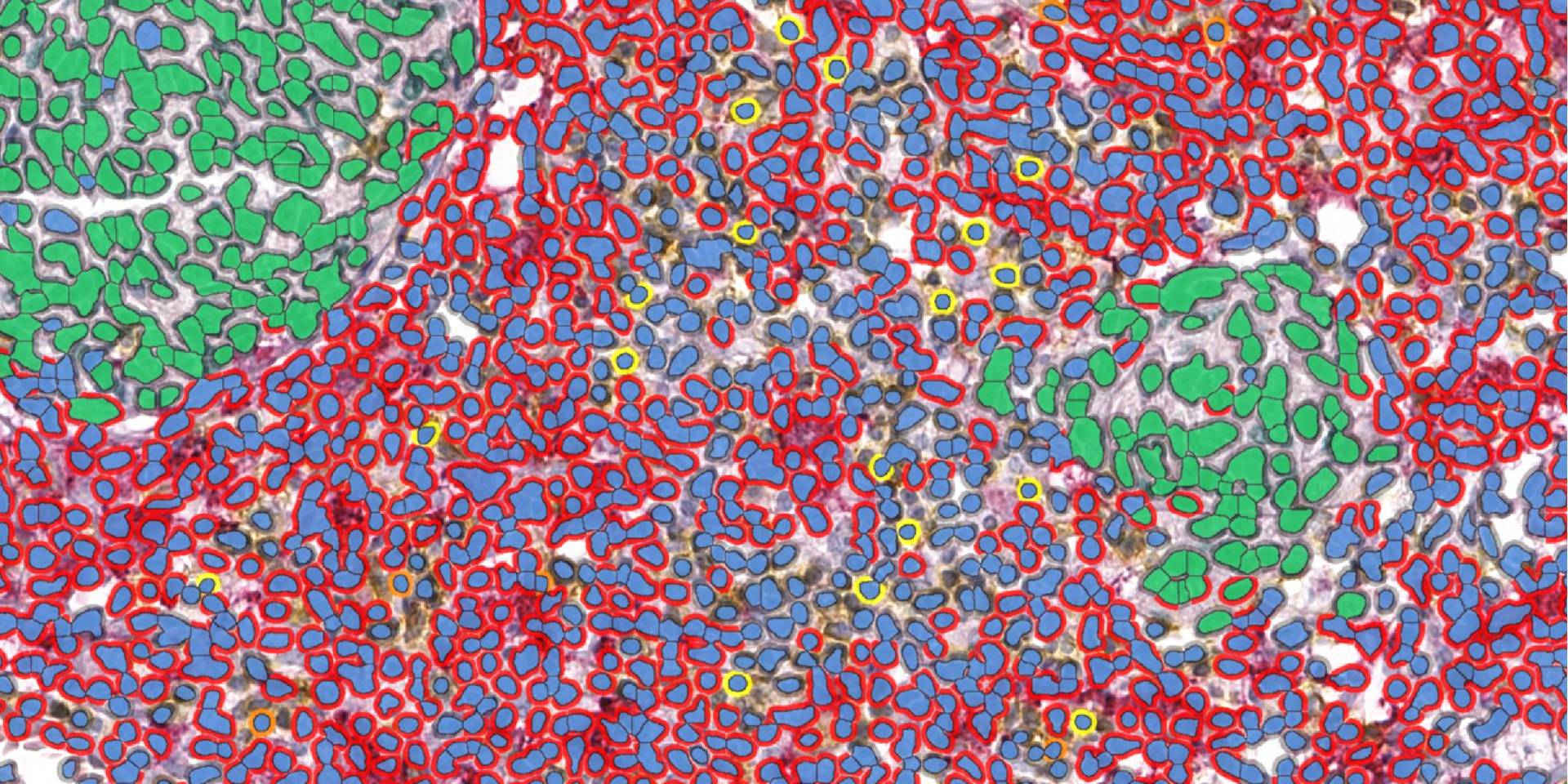
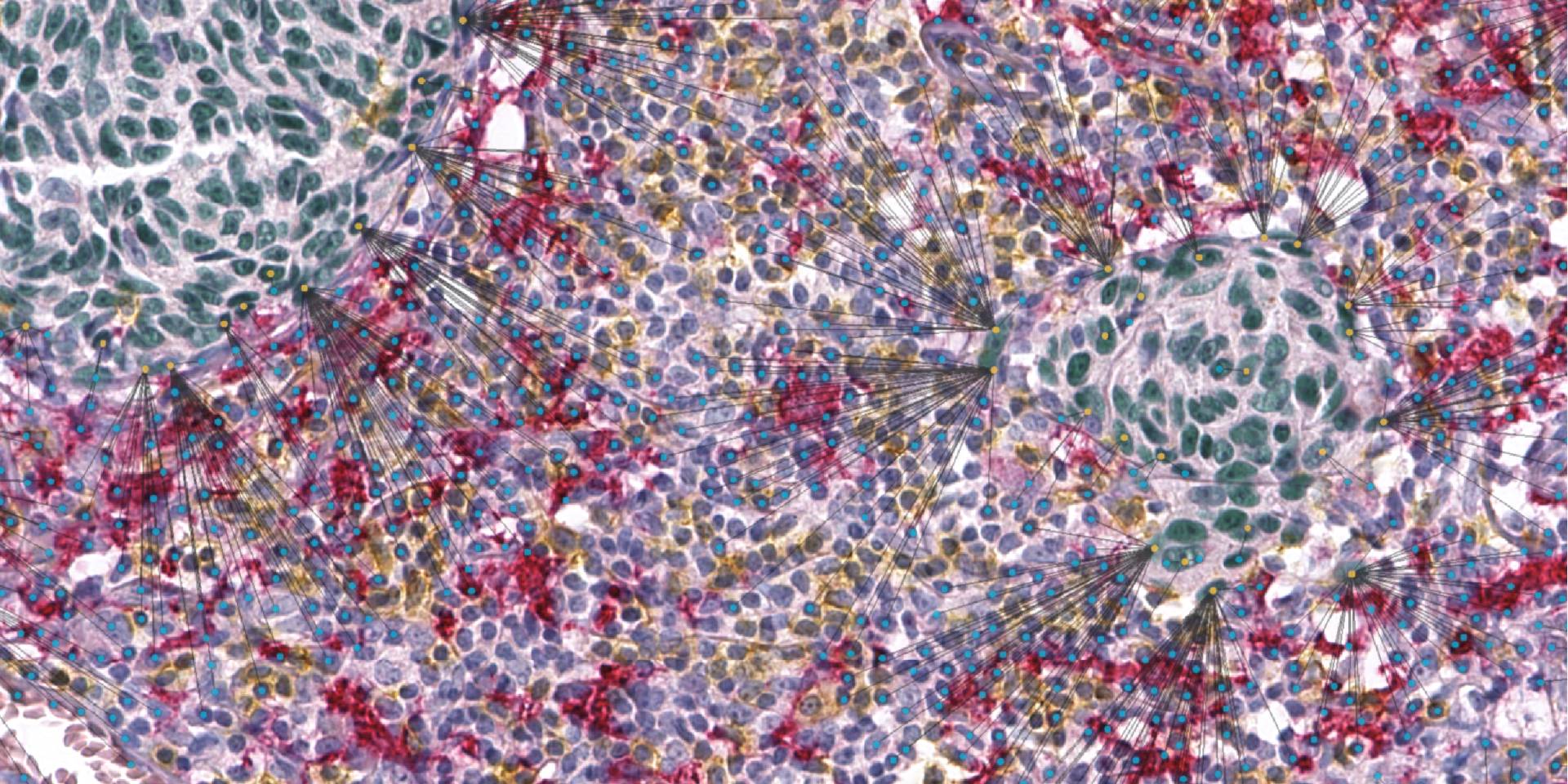
fluorescence up to 9 fluorochromes. - HALO (Indica Labs) software for advanced image analysis on digital scans that can
perform cell counts in brightfield by discriminating up to 5 markers and define relative
distances between cells and tissue borders. - IMARIS software for 3D reconstruction of fluorescence acquisitions on Z-axis multiple
planes. - HALO Link Sharing Platform (Indica Labs), perfectly integrated with the image analysis
modules, capable of supporting different format digital scans from any digital scanner with
the possibility of sharing via Browser Link. - HALO AI (Indica Labs), artificial intelligence module based on Deep Learning, able to
create neural networks for the identification of tissue areas after dedicated training. The
Image Analysis Workstation is equipped with graphic cards with high computing capacity. - Tissue microarray (Alphelys), with tissue arrays design software Version 1.1.
Last update
16.11.2021